Images made with AutoDockFR (ADFR). Click an image to enlarge it.
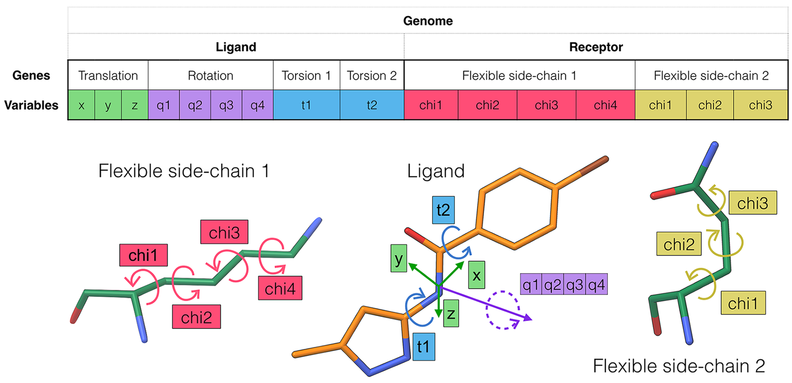
ADFR encodes ligand position, orientation and torsions together with receptor side-chain chi angles in a single genome. Source: Ravindranath et al., PLoS Comput Biol 2015, Fig. 1, CC BY 4.0.
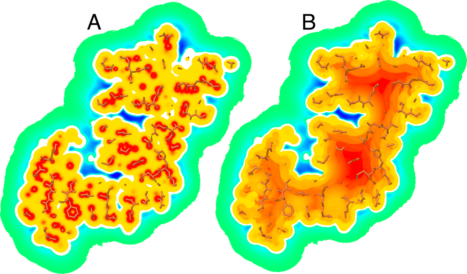
Cross-section of a carbon affinity map before and after adding the gradient that pushes ligand atoms out of the receptor interior. Source: Ravindranath et al., PLoS Comput Biol 2015, Fig. 2, CC BY 4.0.
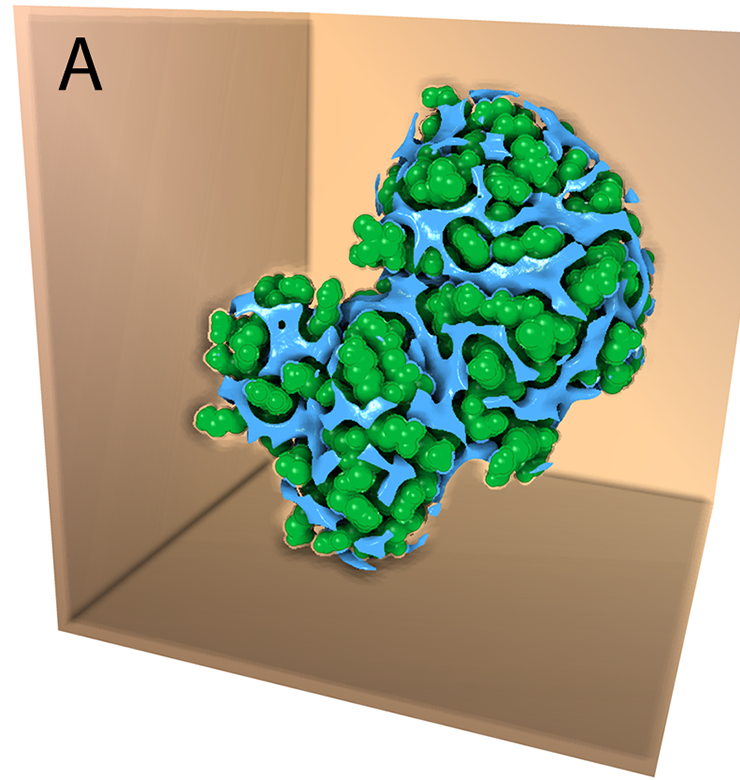
Translational points derived from the affinity maps cover grooves and cavities where a ligand can be placed. Source: Ravindranath et al., PLoS Comput Biol 2015, Fig. 4, CC BY 4.0.
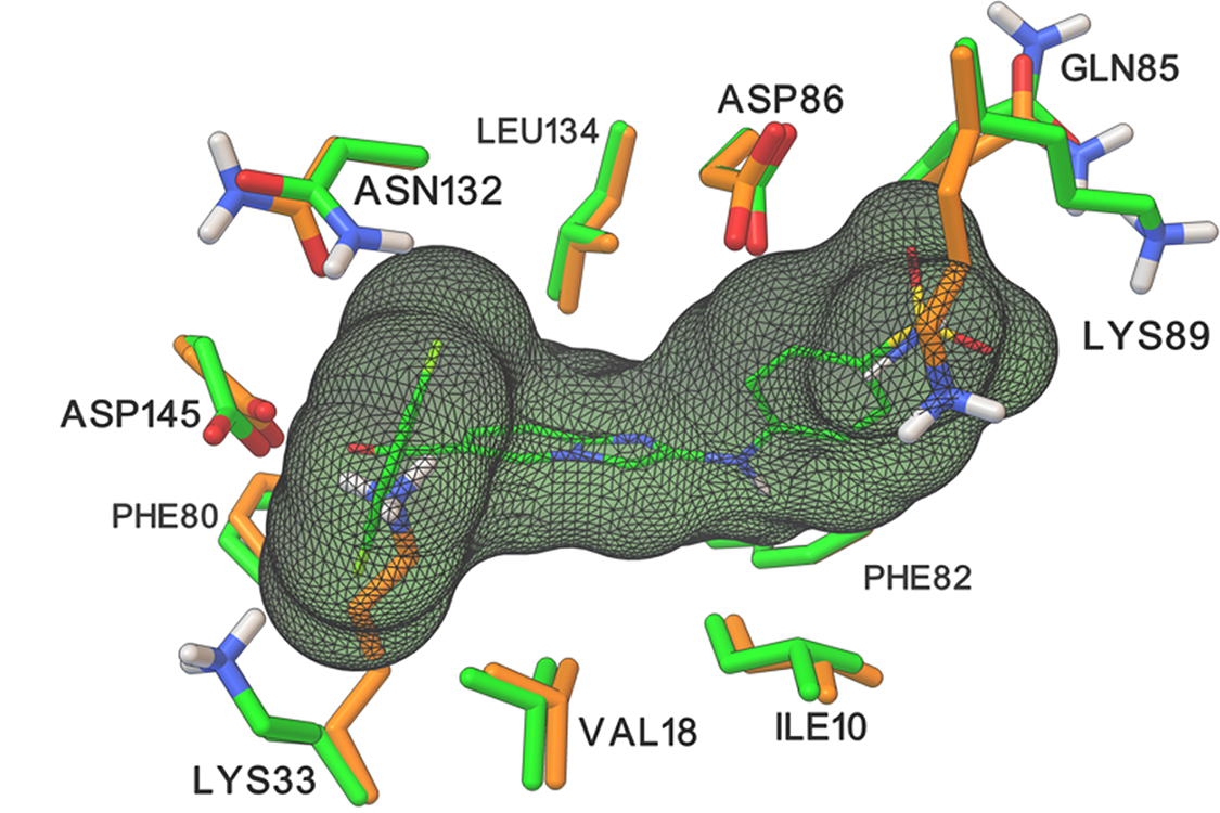
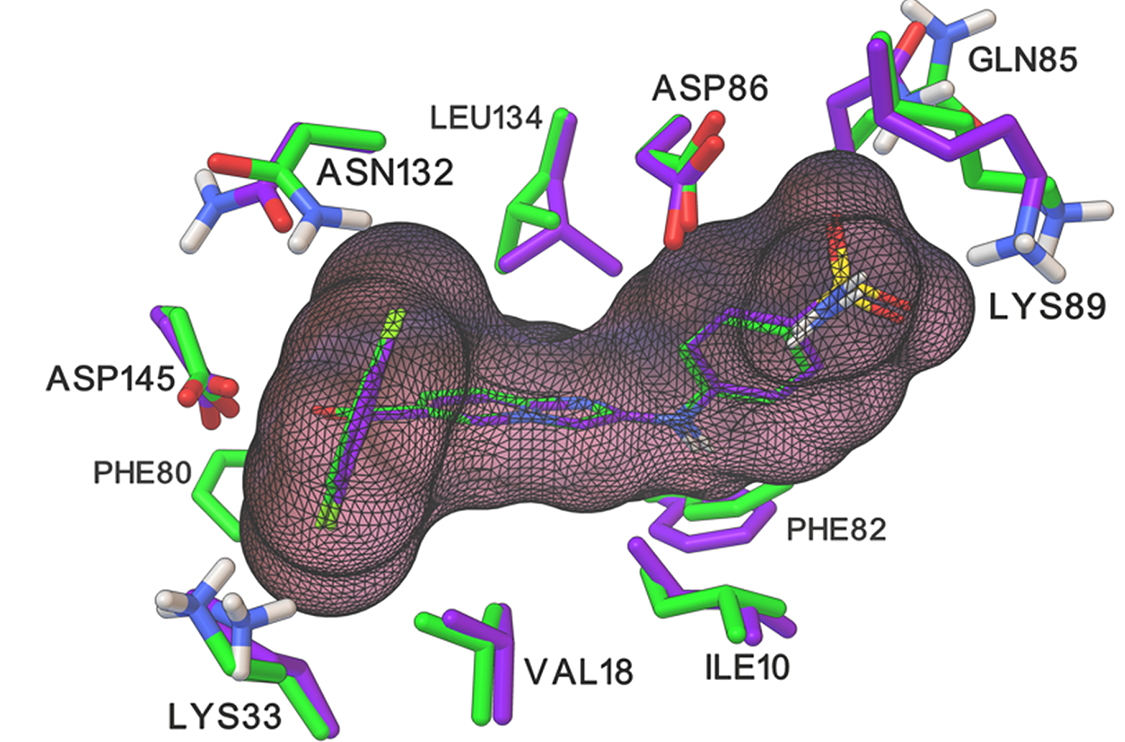
Side-chain conformations of CDK2 binding-site residues differ between the apo and ligand-bound structures. Source: Ravindranath et al., PLoS Comput Biol 2015, Fig. 8, CC BY 4.0.
Docking into apo CDK2 with 12 flexible side chains, ADFR recovers both the ligand pose and the holo side-chain conformations. Source: Ravindranath et al., PLoS Comput Biol 2015, Fig. 8, CC BY 4.0.
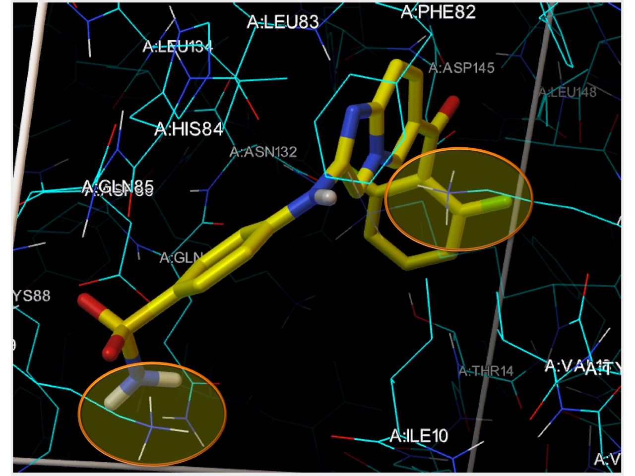
A ligand placed in the apo receptor clashes with Lys33 and Lys89, the side chains ADFR makes flexible. Source: ADFR tutorial, adfr.scripps.edu (2016).